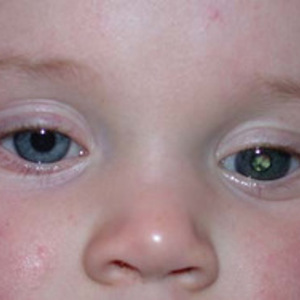
Bilateral RB

Your eye doctor has diagnosed a cancerous tumour known as retinoblastoma in both of your child’s eyes. If there is no history of retinoblastoma in the family, the condition is known as Bilateral Sporadic RB. Treatment of this form of RB depends on the size, number and location of the tumours in each eye.
Enucleation
Usually by the time that Bilateral Sporadic RB is diagnosed, the tumour in one of your child’s eye is very large and your child has been without vision in that eye for some time. Removal or enucleation of this eye is still the commonest form of treatment for the more severely affected eye.
The reasons for performing enucleation in Bilateral Sporadic RB include:
- The tumour inside one of your child’s eye is too large or extensive to safely control with eye saving treatments, such as chemotherapy or radiotherapy.
- There is no hope of preserving vision in this eye.
- Leaving the eye in place may threaten the life of your child.
The thought of your child needing to have an eye removed is a devastating experience for any parent. Under the circumstances, it is important for parents to try and concentrate on a few key points:
- Treatment of retinoblastoma is the most successful of all the childhood cancers. In Bilateral Sporadic RB, removal of the more severely affected eye will improve the chance of curing the condition in your child.
- By the time a child is diagnosed with Bilateral Sporadic RB, they have often been without useful vision for some time in the eye that needs removal. You may not have noticed any difference in your child’s visual behaviour prior to the diagnosis and removal of an eye will not suddenly make life more difficult for them.
- Many children who have had one eye removed can lead a completely normal life. If the vision in the other eye is not severely affected, your child should have no difficulty with schooling, may still be able to drive a car, and will almost certainly be able to enjoy a fulfilling and successful career.
Please go to “Enucleation” for details regarding the operation, post-operative care and the artificial eye.
Chemotherapy
After enucleation, chemotherapy is now the most important initial or primary treatment for Bilateral Sporadic RB. It is commonly used for treatment of the less severely affected eye if the other eye has been removed, but may also be used to treat both eyes if they are equally affected.
 The chemotherapy regimen usually takes the form of eight rounds or cycles of “triple therapy” (carboplatin, vincristine and etoposide) over six to eight months. This will be administered by your childhood cancer doctor (paediatric oncologist) who will discuss the details and side effects of treatment with you. Your child will require an EUA of the eyes after every two cycles of chemotherapy to assess the response of the tumours to the treatment.
Prior to commencing chemotherapy, your child will require a lumbar puncture and bone marrow biopsy. They will also require placement of an infusaport, through which the chemotherapy will be given over the duration of their treatment. All of these procedures can hopefully be carried out under the one general anaesthetic. Your paediatric oncologist will discuss the details and risks of all of these procedures with you.
New and Recurrent RB Tumours
About one half of the children treated with chemotherapy will develop further small new tumours despite chemotherapy and about one quarter will have recurrences of the tumours already treated with chemotherapy. In order to pick these tumours up at the earliest possible time, your child will require EUAs until at least the age of five and will need continued examination in the clinic for a few more years after this. Treatment of new and recurrent tumours is usually possible by one of the local treatment methods outlined in the next section. Please go to “Follow Up” for further information regarding follow up of patients with Bilateral Sporadic RB.
Local Treatment Methods
Local treatment methods can be used in addition to chemotherapy to try and reduce the risk of the tumours recurring following chemotherapy. Alternatively, because of the risk of visual loss with any of these methods, they can be held in reserve and used for the treatment of recurrent tumours as well as for any new tumours that may be seen during follow up EUAs.
Cryotherapy Freezing
Cryotherapy Freezing treatment or cryotherapy of new or recurrent RB is a very effective method of controlling small tumours of the retina toward the front of the eye.
Cryotherapy must be performed while your child is asleep under a general anaesthetic. An instrument known as a cryoprobe (see photo above) is placed on the outer surface of the sclera (white of the eye) at the exact location of the tumour inside the eye. The tumour is then destroyed by freezing of the tumour cells with very little damage to the surrounding healthy tissue.
Your child’s eye may be a little red and sore and the eyelids may be a little swollen for a few days after the treatment. You will need to place an antibiotic drop (eg. Chlorsig) and a mild steroid anti-inflammatory drop (eg. Predsol) in your child’s eye three times per day for one week after the treatment. There is a small risk of bleeding into the eye, lifting or detachment of the retina, and loss of eyesight or even blindness after cryotherapy.
Cryotherapy may need to be repeated one month later, particularly when used for new or recurrent tumours.
Diode Laser Thermotherapy Heating
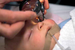
Diode Laser Thermotherapy Heating treatment of new or recurrent RB with an infra-red diode laser is a very effective method for controlling small tumours of the retina toward the back of the eye. This is often known as Transpupillary Thermotherapy (TTT).
TTT must be performed while your child is asleep under a general anaesthetic. The laser beam is directed at the tumour through the dilated pupil using a special lens delivery system (see photo above). Heating of the tumour cells may take several minutes and is often given following a single dose of chemotherapy (carboplatin) administered by infusion prior to the anaesthetic. This is often known as Chemothermotherapy and appears to be more effective than TTT alone.
You will not notice any side effects in your child’s eye following TTT. There is a risk of damage to the eyesight, but only if the central vision area (macula) or main nerve of the eye (optic nerve) are heated by the laser beam.
TTT may need to be repeated one month later, particularly when used for new or recurrent tumours.
Plaque Radiotherapy Radiation
Plaque Radiotherapy Radiation treatment of new or recurrent RB with a small curved radioactive disc (plaque radiotherapy) is very effective for controlling larger tumours of the retina toward the front of the eye.
Plaque radiotherapy must be performed while your child is asleep under a general anaesthetic. The plaque is sutured to the outer surface of the sclera (white of the eye) at the exact location of the tumour inside the eye. The plaque is inserted at one operation and removed at a second operation within two or three days. The tumour is destroyed by irradiation of the tumour cells whilst the plaque is in place. The most common form of plaque used contains a radioactive substance known as Ruthenium-106. The design of the plaque ensures that there is minimal damage to the surrounding healthy tissue.
Your child’s eye will be a little red and sore and the eyelids may be a little swollen for a few days after the treatment. You will need to place an antibiotic drop (eg. Chlorsig) and a mild steroid anti-inflammatory drop (eg. Predsol) in your child’s eye three times per day for two weeks after the treatment.
There is a small risk of loss of vision in the future due to radiation damage to the healthy retinal tissues of the eye (radiation retinopathy) and a risk of clouding of the lens of the eye (cataract).
Your eye doctor will further discuss the details and risks of this form of treatment with you if the need arises.
External Beam Radiotherapy
In recent years chemotherapy has replaced external beam radiotherapy (EBXRT) as the first line of treatment for Bilateral Sporadic RB. The main reasons for this were the desire to avoid the severe side effects of this form of radiotherapy on the eye and face and the possible risk of causing new tumours in the bones or tissues around the eye.
EXBRT is now used mainly for the uncommon situation where there are large or severe recurrences in the only eye following the use of chemotherapy, particularly if there is a high risk of damage to the vision from the local treatments noted above. Your eye doctor and radiotherapy doctor (radiation oncologist) will discuss the details and risks of this form of treatment with you if the need arises.
Follow Up
Please go to “Follow Up” for further information regarding ongoing review of patients with Bilateral Sporadic RB.
Second Malignant Tumours
Unfortunately, your child remains at risk of a number of other cancers elsewhere in the body for the rest of their life. They will require ongoing review by their paediatric oncologist throughout their childhood and teen years. Later in life, a yearly visit to their local general practioner would be advisable. Any unusual symptoms should be immediately reported to their doctor.
Gene Testing
Retinoblastoma is due to the presence of errors or mutations in a single pair of genes (RB1 gene) in your child’s genetic make-up or DNA. The RB1 gene normally acts to prevent or suppress tumour growth in the retinal cells.
In Bilateral Sporadic RB, the mutations cause malfunction of the RB1 gene in every cell of your child’s body and so allows for the possible growth of a number of retinoblastoma tumours in the retinal cells of either eye. Please go to “RB Genetics” for further information on the genetic basis of RB and the importance of testing for mutations of the RB1 gene in Bilateral Sporadic RB.
