
Enucleation
Removal of an eye or enucleation is still a very common form of treatment for malignant melanoma involving the inner coats of the eye (Uveal Melanoma)
The reasons for performing enucleation of an eye in uveal melanoma may include:
- The eye contains a tumour that is too large for conservative treatment such as plaque radiotherapy, transpupillary thermotherapy or resection.
- The tumour is extending through the outer coats of the eye.
- The tumour is invading the main nerve (optic nerve) at the back of the eye.
- The tumour is causing raised pressure in the eye.
The Operation

Orbital anatomy
Enucleation is usually performed under a general anaesthetic and you will need to stay in hospital for a couple of nights after the operation. The operation may be performed under local anaesthesia if a general anaesthetic is not advisable.
The eyelids are not cut or altered during the operation. There are six muscles and a large nerve (optic nerve) attached to the eye. These all need to be divided before the eye can be removed.
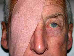
An orbital implant, approximately two centimetres in diameter, is placed into the space that remains after the eye has been removed. The most commonly used implant in adults is made from an artificial coral-like substance known as hydroxyapatite, however an acrylic ball implant may be used. Four of the muscles are reattached to this implant so that the new artificial eye will have the potential for movement. The conjunctival tissue (the thin transparent layer of tissue over the white of a healthy eye) is then closed over the implant. A clear curved plastic disc, called a conformer, is placed in the space between the eyelids and the conjunctiva overlying the implant (socket). A firm bandage is then placed over the area and will remain in place for one and a half to two days after the operation.
Pad on eye following operation
Following the Operation
Following the operation there will inevitably be some discomfort , which will lessen over the subsequent 24 hours. The nursing staff on the ward will make sure that optimum pain relief will keep you as comfortable as possible.
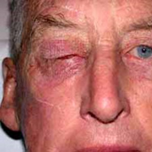
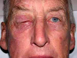
One and a half to two days after the operation the pad will be removed by your eye doctor and you will be able to go home. You will notice that the eyelids will be swollen and perhaps a little bruised, but this will settle rapidly over the next few days. The upper eyelid will remain a liitle droopy until your artificial eye is fiited.
Pad off following operation
You will notice a pinkish-red colour when the eyelids are opened, but will be relieved to see that the appearance is not at all disturbing or offensive – it looks much like the inside of your mouth. You will see the clear plastic conformer sitting in place behind the eyelids. The conformer helps to maintain the normal position of the eyelids and prevent shrinkage of the tissues of the socket before the artificial eye is fitted. It does not need to be touched, but occasionally it may fall out. If this happens, there is no need to panic. Just collect the conformer, clean it with soapy water and bring it in to your eye doctor in an envelope when you next visit. Your doctor will replace the conformer, but there is no urgency and there is no problem if it remains out until this time.
You may have mildly blood stained tears for a few days after the operation, but this is not a concern. If you notice a large amount of fresh blood or if the eyelids become hot, red and swollen, contact your eye doctor immediately.
Medications
You will need to take antibiotic tablet (eg. Keflex) for two weeks following the operation and you will need to place an antibiotic drop (eg. Chlorsig) in the eye three times per day for the same period of time.
Pathology Results
One week following the operation you must return to see your eye doctor to discuss the results of cellular examination of the eye and tumour (pathology).
Orbital Radiotherapy
Radiotherapy to the tissues of the bony cavity that contained the eye (orbit), is carried out if the pathology results show that there is evidence of spread of the tumour through the outer coats of the eye. The aim of radiotherapy is to reduce the chance of the tumour recurring in the orbital tissues. Your eye doctor and radiotherapy doctor (radiation oncologist) will discuss the details of orbital radiotherapy if the need arises, but fortunately the risk is very low.
Artificial Eye
The artificial or prosthetic eye will be made four to six weeks after the enucleation. Please go to “Artificial Eye” for more information.