Dysplastic Lesions
Treatment
Treatment of dysplastic lesions on the surface of the eye depends on a number of factors, including size and location of the lesion. Most of these lesions are located at the border between the conjunctiva (the thin transparent layer of tissue over the “white” of the eye) and the cornea (the clear “window”of the eye), an area known as the limbus.
Small Localised Dysplastic Lesions
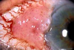
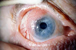
Early Dysplastic Lesion
For very small localised dysplastic lesions, it is usually safe to observe for evidence of growth before recommending surgical removal. The lesion will most likely need to be photographed on a special camera known as a Slit Lamp Camera, so that the photo can be used for future comparisons. You may only need to be observed on a yearly basis regarding this lesion.
If your eye doctor decides to keep your suspicious lesion under observation, it is vital that follow-up appointments are made before leaving after each visit. If you cannot make an appointment or miss a scheduled appointment, then it is important that you ring the rooms to make a new appointment.
Larger Localised Dysplastic Lesions
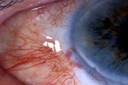
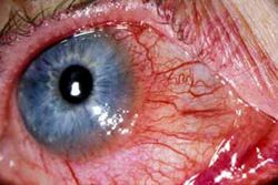
Larger Localised Dysplastic Lesion
For larger localised dysplastic lesions, it is best to completely remove or excise the lesion during a short operation.
Operation
Excision of dysplastic lesions is usually performed under local anaesthesia as a day surgery procedure, and basically involves shaving the lesion off the surface of your eye. This allows for an immediate cellular or pathological diagnosis, in particular to exclude a cancerous lesion, and has the added benefit of near complete clearance of the lesion from the surface of your eye. Freezing treatment (cryotherapy) will be applied to the edge of the surgical wound at the time of the operation, as we know that this reduces the significant risk of future recurrence of the lesion.
Risks of the operation
With any surgical procedure on the eye, there is a very small risk of loss of vision, however the risks are higher if the lesion is left to grow untreated on the eye. There is also a small risk of infection, formation of excessive healing tissue (granuloma) and occasionally the area of excision may remain pinker than the surrounding tissue of the eye.
Following the operation
Following the operation, you will be placed on antibiotic (eg.”Chlorsig”) and anti-inflammatory (eg.”Prednefrine Forte”) drops for at least a couple of weeks. You will be asked to return for review of the wound and to discuss the results of the pathology within one week of your operation.
Pathology
If the pathology result reveals intra-epithelial neoplasia, this means that the dysplasia is confined to the surface layer (cornea or conjunctiva) of your eye, is not invasive, but must be regarded as being pre-cancerous. There is a significant risk that the lesion can recur if no further treatment is given. Your eye doctor will then advise additional or adjunctive treatment with chemotherapy drops known as Mitomycin C to further reduce the risk of recurrence of the lesion. Please go to “Mitomycin C” for further information regarding treatment of dysplasia with these drops.
If the pathology reveals invasive neoplasia (squamous cell carcinoma), this means that the dysplasia is invasive and that there is a significant risk that this cancerous lesion can destroy the eye if no further treatment is given. Your eye doctor will then advise additional or adjunctive treatment with a localised form of radiation treatment known as Beta Radiotherapy to reduce the risk of the lesion invading the eye or the tissues surrounding the eye. Please go to “Beta Radiotherapy” for further information regarding treatment of non-invasive dysplasia with this form of radiotherapy.
Diffuse Dyspalstic Lesions
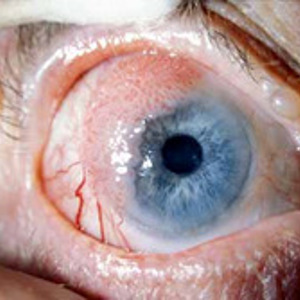
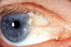
DIffuse Dysapastic Lesion pre surgery
Less commonly, the dysplastic lesion more extensively or diffusely involves the surface of your eye. In this situation you will need to have a small biopsy or biopsies taken from the lesion to determine its pathological nature. This is usually done as a day surgery procedure under local anaesthesia.
Most commonly the lesion is pre-cancerous intra-epithelial neoplasia (see above). If this is the case, further surgery may be avoided and the lesion treated with chemotherapy drops alone. Please go to “Mitomycin C” for further information regarding treatment of diffuse dysplasia with these drops.
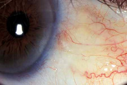
Small dysplasia before and after excision
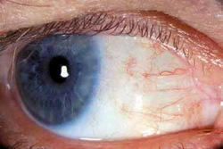
Large dysplasia before and after excision